Nanobubbles in oncology research
What have laboratory and animal studies reported about nanobubbles for tumor oxygenation, drug delivery and imaging?
On this page
Tumor hypoxia and oxygen delivery
Researchers have used oxygen nanobubbles to raise oxygen levels in hypoxic tumor cells and tumors, a step studied as a way to overcome treatment resistance Khan et al., 2018; Song et al., 2020.
Why hypoxia matters in tumors
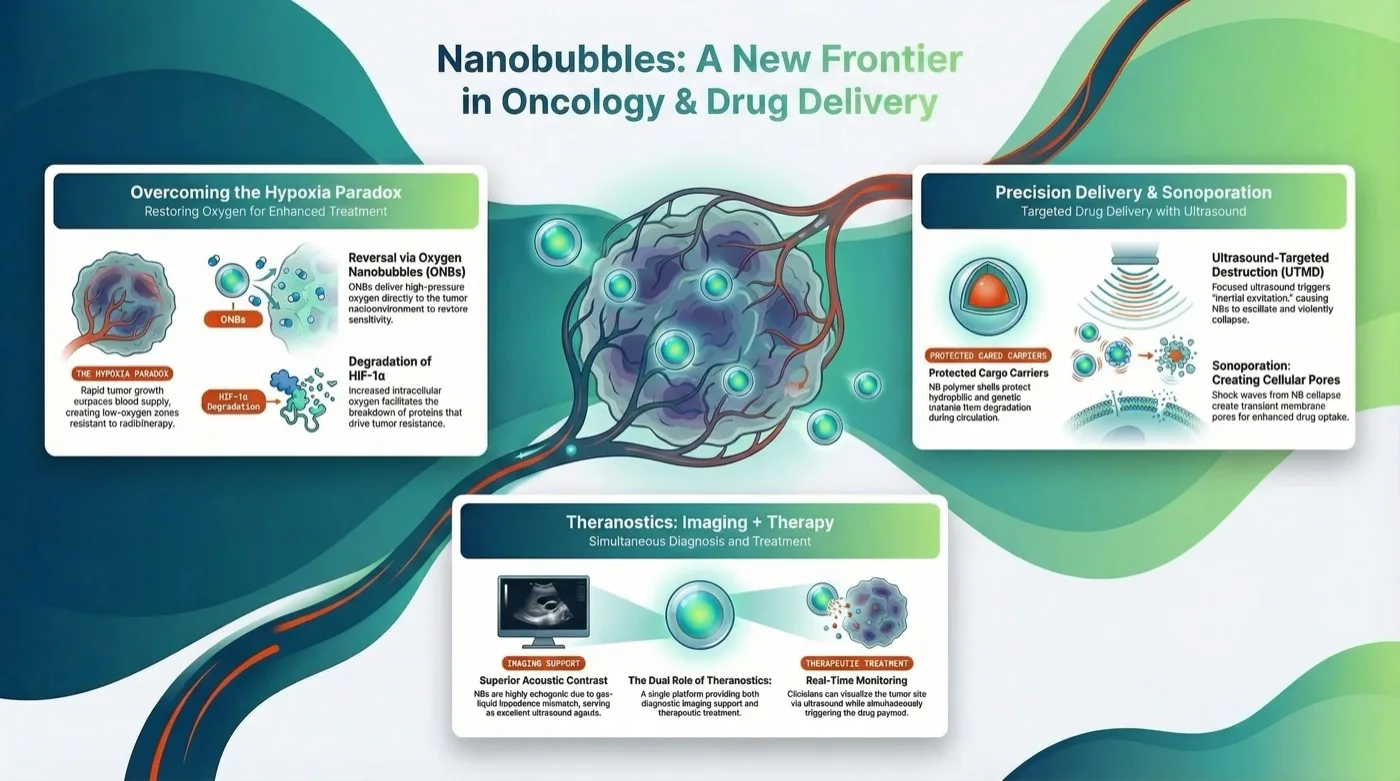
Fast-growing solid tumors often outgrow their blood supply. Their vessels are irregular and leaky, and parts of the tumor become severely short of oxygen (). This hypoxia makes tumors harder to treat. Radiotherapy relies on oxygen to "fix" the DNA damage caused by free radicals; without oxygen, much of that damage can be repaired and the cells survive. Hypoxia also slows cell division, which blunts chemotherapies that target dividing cells, and it favors more aggressive cell types that are more likely to spread Khan et al., 2018; Song et al., 2020.
HIF-1α and oxygen nanobubbles in cell studies
Under low oxygen, the protein hypoxia-inducible factor 1-alpha (HIF-1α) becomes stable, moves into the nucleus and switches on genes linked to cell survival, new blood-vessel growth (angiogenesis) and spread (metastasis). When oxygen returns, HIF-1α is modified (hydroxylated) and broken down.
Khan et al. made lipid-shelled oxygen nanobubbles by sonication and tested them on MDA-MB-231 breast cancer cells under hypoxic conditions. As oxygen was released, intracellular oxygen rose and HIF-1α was hydroxylated and degraded. The researchers reported a significant reduction in HIF-1α expression and a matching fall in hypoxia-associated fluorescence markers (Image-iT) Khan et al., 2018. Reversing hypoxia in this way is the proposed route to making cells more responsive to treatment.
Oxygen delivery in vivo
Song et al. used lipid-coated biogenic gas vesicles (lipid-GVs) to carry oxygen. After tail-vein injection, oxy-hemoglobin levels in subcutaneous tumors rose within 15 minutes, and the researchers reported that tumor hypoxia eased and HIF-1α downstream pathways were downregulated Song et al., 2020.
Nanobubbles as drug and gene carriers
Laboratory studies and reviews describe nanobubbles as carriers for drugs and genetic material, with a shell that protects the cargo Cavalli et al., 2015; Jin et al., 2022.
Cavalli et al. developed chitosan-shelled nanobubbles with a perfluoropentane core, sometimes called nanodroplets. They loaded them with a corticosteroid, prednisolone phosphate, or coupled them with an MRI contrast agent, Gd-DOTP. The polymer shell protects the cargo from degradation. Particles of this size may leave the bloodstream through leaky tumor vessels, a mechanism known as the enhanced permeability and retention (EPR) effect Cavalli et al., 2015.
Reviews also describe nanobubbles as non-viral carriers for genetic material such as siRNA and plasmid DNA. The shell may protect the material from enzymes in the blood, and the bubbles can be targeted to specific tissues Jin et al., 2022; Wu et al., 2021. These are review-level descriptions of a research field, not results from a single controlled study.
Ultrasound-triggered release and sonoporation
In laboratory work, ultrasound increased drug release from nanobubbles and was used to open transient pores in cell membranes Cavalli et al., 2015; Chang et al., 2016. The approach is known as ultrasound-targeted microbubble destruction (UTMD).
The idea is to release the drug where it is needed. Once bubbles have gathered at a tumor, focused ultrasound makes them oscillate and then collapse (inertial cavitation). The collapse produces micro-jets and shock waves that disrupt nearby cell membranes and open transient pores, a process called sonoporation.
This on-demand release is studied as a way to raise the uptake of drugs such as doxorubicin or paclitaxel, which cross cell membranes and the blood-brain barrier poorly. Cavalli et al. showed that ultrasound stimulation of chitosan nanobubbles markedly increased the rate of drug release compared with unstimulated samples. The goal is a high local drug concentration with less exposure elsewhere in the body Cavalli et al., 2015; Chang et al., 2016. Cavalli et al.'s release tests were in vitro, so they could not show lower systemic toxicity.
Imaging and delivery combined
Because the gas core reflects ultrasound strongly, nanobubbles are echogenic and can act as ultrasound contrast agents. Researchers have combined this with drug or oxygen loading in "theranostic" designs, which aim to image and treat with the same particle Song et al., 2020; Cavalli et al., 2015.
Song et al. used their lipid-coated gas vesicles for both roles. Their acoustic properties gave high-contrast ultrasound images to locate the tumor and follow perfusion. They also carried oxygen to support photodynamic therapy (PDT), which uses light to generate (reactive oxygen species) in the tumor. With the oxygen-loaded vesicles present, ROS generation under laser irradiation increased, and tumor growth was inhibited more than with PDT alone Song et al., 2020.
Cavalli et al. showed that chitosan nanobubbles loaded with Gd-DOTP, a gadolinium-based contrast agent, could be seen by magnetic resonance imaging (MRI) while also responding to ultrasound as drug carriers. The authors presented this as a route to monitoring by more than one imaging method Cavalli et al., 2015.
Limits and open questions
This is early-stage research. The findings come from cancer cell lines, laboratory release tests and animal tumor models; no human clinical trial is cited, and results in cells or animals often do not carry over to patients.
Each finding rests on one or two studies. The lesson does not give sample sizes, doses or treatment durations, and the drug-carrier and gene-delivery material comes partly from reviews rather than primary experiments.
The oxygen-delivery studies used specialized particles, lipid-shelled oxygen nanobubbles and biogenic gas vesicles, prepared in the laboratory and given by injection or added to cell cultures. They say nothing about drinking, bathing in or otherwise using oxygenated water. Safety, dosing and long-term effects in people are not established by the work summarized here.
References
- Khan, M. S., Hwang, J., Seo, Y., et al. (2018). Engineering oxygen nanobubbles for the effective reversal of hypoxia. Artificial Cells, Nanomedicine, and Biotechnology, 46, 318-327. https://doi.org/10.1080/21691401.2018.1492420 ↩
- Song, L., Wang, G., Hou, X., et al. (2020). Biogenic nanobubbles for effective oxygen delivery and enhanced photodynamic therapy of cancer. Acta Biomaterialia, 108, 313-325. https://doi.org/10.1016/j.actbio.2020.03.034 ↩
- Cavalli, R., Argenziano, M., Vigna, E., et al. (2015). Preparation and in vitro characterization of chitosan nanobubbles as theranostic agents. Colloids and Surfaces B: Biointerfaces, 129, 39-46. https://doi.org/10.1016/j.colsurfb.2015.03.023 ↩
- Jin, J., Yang, L., Chen, F., et al. (2022). Drug delivery system based on nanobubbles. Interdisciplinary Materials, 1, 471-494. https://doi.org/10.1002/idm2.12050 ↩
- Wu, R., Yang, X., Li, X., et al. (2021). Nanobubbles for tumors: Imaging and drug carriers. Journal of Drug Delivery Science and Technology, 65, 102749. https://doi.org/10.1016/j.jddst.2021.102749 ↩
- Chang, S., Si, T., Zhang, S., et al. (2016). Ultrasound mediated destruction of multifunctional microbubbles for image guided delivery of oxygen and drugs. Ultrasonics Sonochemistry, 28, 31-38. https://doi.org/10.1016/j.ultsonch.2015.06.024 ↩