Characterization and measurement
How do you confirm that a liquid contains nanobubbles, and how are they counted?
On this page
What the eye can and cannot tell you
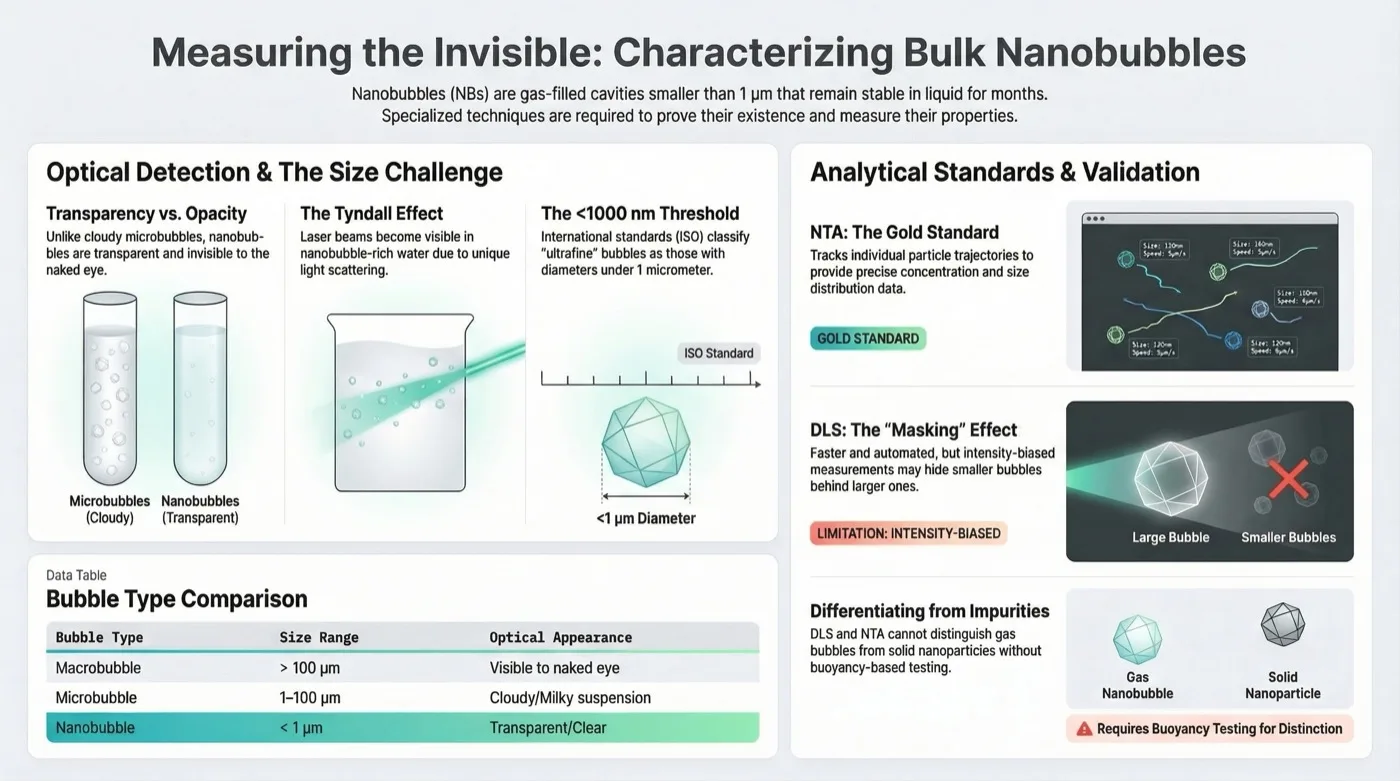
Turbidity shows microbubbles, not nanobubbles. When generated, microbubbles (typically 10–50 µm) scatter enough light to make water look milky or cloudy; because they rise quickly and burst at the surface, the milkiness fades soon after Agarwal et al., 2011.
Nanobubbles are much smaller, with reported sizes from under 200 nm up to 1 µm, and have negligible buoyancy. They do not scatter enough light to make water opaque, so a nanobubble suspension looks transparent even at high concentrations Etchepare et al., 2017; Babu and Amamcharla, 2023. Clear water is therefore consistent with nanobubbles, but it does not confirm them; that takes instruments.
Nanoparticle tracking analysis: tracked particles per mL
(NTA) is the preferred method in the cited studies because it counts as well as sizes. A laser beam illuminates particles in a liquid sample, and a microscope with a high-sensitivity camera records the light each one scatters. Software follows the Brownian motion of every particle frame by frame and, using the Stokes–Einstein equation, converts its diffusion speed into a hydrodynamic diameter: smaller particles move faster Nirmalkar et al., 2018; Azevedo et al., 2016.
NTA reports a number concentration, in tracked particles per mL, alongside the size distribution. It resolves individual particles even in polydisperse samples, where ensemble methods bias results toward larger particles. Studies of nanobubble existence and stability have used it for this reason Alheshibri et al., 2016; Ushikubo et al., 2010; Azevedo et al., 2019.
What NTA counts is scattering objects, not bubbles specifically. A gas nanobubble, a solid nanoparticle and an oil droplet of the same size can all be tracked, so an NTA result is a count of particles until other evidence shows they are gas-filled. That is why the studies above pair NTA with the tests described below Nirmalkar et al., 2018.
Dynamic light scattering: size, not concentration
(DLS) estimates an average size from fluctuations in scattered light intensity caused by Brownian motion. It is widely used because it is easy to operate and covers sizes from below a nanometer up to the micron range Gurung et al., 2016; Li and Zhang, 2022.
Its main limitation for nanobubbles is weighting. Scattered intensity is proportional to the sixth power of diameter (d⁶), so a few large microbubbles or contaminants can mask the signal of millions of nanobubbles and push the reported mean size upward. DLS also cannot provide the number concentration of bubbles, a key parameter for evaluating a nanobubble generator Nirmalkar et al., 2018; Li and Zhang, 2022.
Telling bubbles from impurities
Researchers use stress tests to separate gas-filled nanobubbles from solid nanoparticles or oil droplets, because the counting instruments alone cannot.
- Compression. Nanobubbles are compressible: under external pressure they shrink or dissolve, whereas solid nanoparticles keep their size. The response is tracked with DLS or NTA under pressure Alheshibri and Craig, 2018; Ahmed et al., 2018.
- Freeze–thaw. Freezing destroys nanobubbles, while solid contaminants remain detectable. A large drop in particle count after thawing indicated that the original population was mostly nanobubbles Nirmalkar et al., 2018; Li et al., 2021.
- Resonant mass measurement. RMM separates particles by buoyancy. Nanobubbles have negligible mass and are positively buoyant in liquid, whereas solid particles are negatively buoyant, which distinguishes gas from solid phases Alheshibri and Craig, 2018; Azevedo et al., 2019.
Controls before calling particles bubbles
A particle count becomes a bubble count only when controls rule out the other explanations. Alongside the stress tests above, three controls address the most common ones:
- Filtered-water blank. Run the water used to make the sample, filtered and without gas treatment, through the same instrument and settings. Its count is the background that treatment has to exceed.
- Degassed control. Measure a sample processed the same way with the gas removed or omitted. Particles that appear regardless of gas are unlikely to be bubbles.
- Stability over time. Measure the same sample repeatedly after generation, and the controls the same way. A single reading cannot show whether the population is stable, growing or disappearing.
Results should then be reported as measured: particles per mL, with the instrument, settings, temperature and any dilution stated.
What this means in practice
In practice, a site or pilot that wants to know whether its water carries nanobubbles should plan the measurement before sampling. Take samples at the generator outlet and at the point of use, and have them analyzed by NTA together with a filtered-water blank and a degassed or untreated control from the same source water.
Ask the laboratory to report concentration as particles per mL, as measured, with the dilution, temperature and instrument settings recorded, and to repeat the measurement on stored samples over time. Where the result will be used to make a decision, add one stress test (freeze–thaw, compression or RMM) to support calling the particles bubbles.
Keep (dissolved oxygen, in mg/L) as a separate record: a DO probe measures dissolved gas, not bubbles, and neither measurement substitutes for the other.
Limits and open questions
The methods here are described from laboratory studies and review articles. The cited work does not report how often each control or stress test is used, or how well each performs in water with high background particle loads, such as irrigation, pond or process water.
Each discrimination test addresses one alternative explanation, such as solid particles or oil droplets; the lesson cites no single test that settles all of them.
The sizes and counts discussed are reported from specific instruments and conditions, and NTA results depend on the settings used, so results from different laboratories may not compare directly.
Questions
Does nanoparticle tracking analysis count nanobubbles?
It counts tracked particles per mL. NTA follows the Brownian motion of anything that scatters laser light, so gas bubbles, solid nanoparticles and oil droplets can all appear in the count. A result supports calling those particles bubbles only with controls such as a filtered-water blank, a degassed control and a stress test.
Why not use dynamic light scattering to measure nanobubbles?
DLS reports an average size, not a count, and scattered intensity scales with the sixth power of diameter. A few large microbubbles or contaminants can therefore mask millions of nanobubbles and shift the reported size upward. It remains useful as a quick size check alongside counting methods.
How can you tell a nanobubble from a solid nanoparticle?
Researchers use stress tests. Under pressure, bubbles shrink or dissolve while solids keep their size; freezing and thawing destroys bubbles but not solids; and resonant mass measurement separates positively buoyant bubbles from negatively buoyant solids. A large count drop after such a test indicates bubbles.
References
- Nirmalkar, N., Pacek, A. W., Barigou, M. (2018). On the Existence and Stability of Bulk Nanobubbles. Langmuir, 34, 10964-10973. https://doi.org/10.1021/acs.langmuir.8b01163 ↩
- Azevedo, A., Etchepare, R., Calgaroto, S., et al. (2016). Aqueous dispersions of nanobubbles: Generation, properties and features. Minerals Engineering, 94, 29-37. https://doi.org/10.1016/j.mineng.2016.05.001 ↩
- Li, C., Zhang, H. (2022). A review of bulk nanobubbles and their roles in flotation of fine particles. Powder Technology, 395, 618-633. https://doi.org/10.1016/j.powtec.2021.10.004 ↩
- Agarwal, A., Ng, W. J., Liu, Y. (2011). Principle and applications of microbubble and nanobubble technology for water treatment. Chemosphere, 84, 1175-1180. https://doi.org/10.1016/j.chemosphere.2011.05.054 ↩
- Babu, K. S., Amamcharla, J. K. (2022). Generation methods, stability, detection techniques, and applications of bulk nanobubbles in agro-food industries: a review and future perspective. Critical Reviews in Food Science and Nutrition, 63, 9262-9281. https://doi.org/10.1080/10408398.2022.2067119 ↩
- Li, T., Cui, Z., Sun, J., et al. (2021). Generation of Bulk Nanobubbles by Self-Developed Venturi-Type Circulation Hydrodynamic Cavitation Device. Langmuir, 37, 12952-12960. https://doi.org/10.1021/acs.langmuir.1c02010 ↩
- Alheshibri, M., Craig, V. S. J. (2018). Differentiating between Nanoparticles and Nanobubbles by Evaluation of the Compressibility and Density of Nanoparticles. The Journal of Physical Chemistry C, 122, 21998-22007. https://doi.org/10.1021/acs.jpcc.8b07174 ↩
- Azevedo, A., Oliveira, H., Rubio, J. (2019). Bulk nanobubbles in the mineral and environmental areas: Updating research and applications. Advances in Colloid and Interface Science, 271, 101992. https://doi.org/10.1016/j.cis.2019.101992 ↩
- Etchepare, R., Oliveira, H., Azevedo, A., et al. (2017). Separation of emulsified crude oil in saline water by dissolved air flotation with micro and nanobubbles. Separation and Purification Technology, 186, 326-332. https://doi.org/10.1016/j.seppur.2017.06.007 ↩
- Alheshibri, M., Qian, J., Jehannin, M., et al. (2016). A History of Nanobubbles. Langmuir, 32, 11086-11100. https://doi.org/10.1021/acs.langmuir.6b02489 ↩
- Ushikubo, F. Y., Furukawa, T., Nakagawa, R., et al. (2010). Evidence of the existence and the stability of nano-bubbles in water. Colloids and Surfaces A: Physicochemical and Engineering Aspects, 361, 31-37. https://doi.org/10.1016/j.colsurfa.2010.03.005 ↩
- Gurung, A., Dahl, O., Jansson, K. (2016). The fundamental phenomena of nanobubbles and their behavior in wastewater treatment technologies. Geosystem Engineering, 19, 133-142. https://doi.org/10.1080/12269328.2016.1153987 ↩
- Ahmed, A. K. A., Sun, C., Hua, L., et al. (2018). Generation of nanobubbles by ceramic membrane filters: The dependence of bubble size and zeta potential on surface coating, pore size and injected gas pressure. Chemosphere, 203, 327-335. https://doi.org/10.1016/j.chemosphere.2018.03.157 ↩